[See
ref 13,
ref 16,
ref 17
]
The experimental setup created a microscopic light source out of a single,
small latex beads by aiming a narrow beam of near-infrared light at it. The beam
intensity was pulsating at rates of 1 per second.
The plastic bead scattered the light towards cells nearby, but released no
chemicals that could attract the cells. The heating of the bead on the order of 1/10,000th of a degree
was negligible.
The cells were kept in a live cell chamber with careful
temperature and pH control. In order to be able to see the cells in the microscope the entire chamber
was illuminated with low intensity, visible light of 600 [nm]. The behavior
of the test cell was recorded by video and infrared sensitive CCD cameras.
Note: In the recordings shown here and in other sections the phase contrast images of the beads
appear white. The irradiated bead is surrounded by a halo of light because the
camera is looking directly into the infrared beam. The scattered intensity received by
the cells is approximately 1/1000th of the intensity seen by the camera.
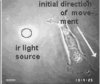
Extension of surface projections towards the pulsating light sources.
We recorded more than 800 cells and found that a statistically highly significant percentage of the cells extended
new pseudopodia towards the plastic beads if they scattered near-infrared light. Often, they displayed
very unusual motile behavior, like the cell shown below which turned 180° in order to reach the
light source.
(The illustration is animated.Click here for a minimal strip of frames.)
The figure below shows the percentage of cells that removed the light scattering particle as a function of wavelength.
The most 'attractive' wavelength was between 800 and 900 [nm].

Absence of heat effects:No convective currents near light spot.
The interpretation that the test cells are capable to detect the light sources at a distance may meet a major objection: The infrared beam may heat the medium as it traverses the observation chamber. The heated medium will rise and thus generate convective currents that flow continuously toward or away from the particle. Consequently, it may not have been the light which guided the cell extensions to the light sources but by the direction of these convective currents. However, we recorded several hours of video sequences which showed that small latex particles inside the chamber near the infrared beam perform normal Brownian motion without any directional component to indicate convective currents.
Absence of chemotactic gradient:Particle pick-up in spite of medium stream towards the particle.
Another objection may claim that the beam produces chemical changes in the medium or at the irradiated particle which set up a chemo-attractive gradient for the surrounding cells. Therefore, we let medium flow through the chamber (speed appr. 2.7 mm/s) and selected situations where the medium streamed from the cell towards the light spot. If there was a chemo-attractive gradient, its material should be driven away from the cell and the cell should not be attracted to the light spot. Nevertheless, the several test cells moved towards the light spot.
Significance for cell intelligence:
It would be strange enough if cells detect near infrared light,
but why does it need to pulsate to be detected?
The photon energy of near-infrared light is too low for photochemical effects on
molecular bonds. Only solid state devices may have energy band gaps that provide
resonance absorption for such photons. As cells are able to detect this light
they must have developed special receptor molecules such as bacterio-chlorophyll
which absorbs near infrared light for the photosynthesis of purple bacteria. One may also
argue that cells have developed special methods to detect signal pulsations in order to
improve the signal-to-noise ratio of
the signals in the midst of violent thermal noise. There are also reasons to suspect that
cells detect pulsating signals in order to improve the angular resolution.
Therefore, it appears likely that cells contain a sophisticated machinery to detect
pulses of a photochemically inactive form of light which is not emitted by any of the
inanimate objects around them. It begs the question whether cells use these
light pulses for communication.