CELL INTELLIGENCE

Guenter Albrecht-Buehler, Ph.D.
Fellow, Institute for Advanced Studies, Berlin
Robert Laughlin Rea Professor of Cell Biology
Northwestern University Medical School, Chicago

SUMMARY.

MINDSET

Depending on the direction in which one reads the next sentence, intelligence is a fractal property or/and an emergent property: ...Intelligent ecologies contain intelligent populations,which contain intelligent organisms, which contain intelligent cells, which contain intelligent compartments, which contain...and so forth.

MAIN HYPOTHESIS

A. Cells control the movement of every part of their body.

Cell movement is not random.. The cortex consists of autonomous domains ('microplasts') whose movement is controlled by a control center (centrosome). Microtubules mediate between the controll center and the autonomous domains.

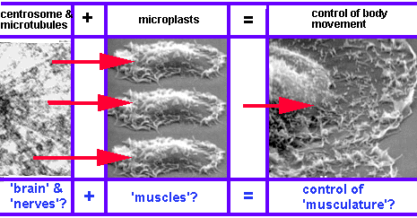
B. The control center detects objects and other cells objects by pulsating near-infrared signals.

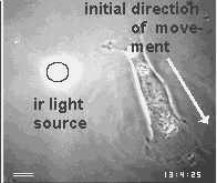
Cell have 'eyes' in the form of centrioles.. They are able to detect near infrared signals and steer the cell movements towards their source.

APPROACH

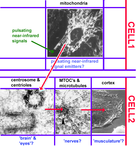
For the past 2 decades I have applied essentially two lines of reasoning to examine whether cells are intelligent. They can be summarized in the following statements:

A. If cells can measure space and time, they must be able to derive abstract data from physical signals.

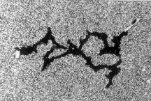
Space and time are not physical objects with which cells could interact, but they are the pre-condition of all physical objects. If cells can measure space and time variables such as angles, distances, curvatures or durations, they must have derived these abstract quantities from the physical objects of their environment. Chapter 2 will use the apparent symmetry and identity between the branches of the phagokinetic tracks of dividing cells (an example is shown below) to argue that cells are programmed to measure angles and time durations.

(The illustration is animated.Click here for a minimal strip of frames.)

B. If cells have eyes, they must be able to order and integrate countless signals.

Images are the ordered set of a huge number of individual data. If cells are capable of generating an image of their environment and react to it, they must be able to order a large number of signals and integrate them into a response action. Chapter 3 will present the evidence that cells use centrioles to 'see' all objects around them that emit or scatter near infrared light. The figure below shows one of the examples of this amazing ability of cells.

(The illustration is animated.Click here for a minimal strip of frames.)

Introduction.

An operational definition of the intelligent cell.

First a disclaimer. My work over the past 2 decades did not intend to join the ongoing efforts of philosophers, logicians and computer scientists to find a universal definition of intelligence. On the contrary, it did not question the common assumption that everybody can tell a mindless, mechanical gadget from an intelligent machine, and proceeded to ask which of the two categories apply to a living cell. Clearly, there are many different levels of intelligence, but I believe that most people consider a machine mechanical and mindless if its actions either do not seem to respond to signals or else always show the same set of reactions. On the other hand, we expect an intelligent machine to responds to signals in a large variety of ways, especially if the signals are unforeseeable, and if its responses offer solutions to problems which were transmitted by the signals. Usually, this means that the intelligent machine contains at least 2 different machines, one which it mindless and carries out some mechanical labor while the other collects and processes signals and controls the action of the first.

Therefore, I went ahead using the following operational definition of an intelligent cell. An intelligent cell contains a compartment which is capable of collecting and integrating a variety of physically different and unforeseeable signals as the basis of problem-solving decisions.

Are there reasons to think that cells are intelligent?

The prevailing wisdom of modern biology has it that cells are immensely complex, but rigidly operating chemical machines that derive their operating instructions internally from their genes and externally from chemicals and electrical signals emitted rigidly by other cells. Unable to believe that any machine can be designed that contains an instruction library which anticipates all the mishaps and glitches of a billion years of evolution without crashing over and over again, I began more than 2 decades ago to search for signs that the cell was actually a 'smart' machine. In other words, I looked for experimental evidence that cells contained a signal integration system that allowed them to sense, weigh and process huge numbers of signals from outside and inside their bodies and to make decisions on their own.

One of the reasons for my disbelief was the sheer size of the organisms that cells built out of themselves. Organism like us are 30,000 times larger in length and and we are not the biggest. Keep in mind that the cells of a gnat are not smaller than the cells of a whale. The whale just contains a lot more cells. How could they build such a huge range of organisms without the ability to override any permanent instructions in order to solve unforeseeable problems on its own?

Anybody who ever built anything bigger than himself knows what frightening surprises it can bring to exceed the dimensions of actions and objects with which we are familiar. The Sears Tower in Chicago is 'only' 300 times larger than the body of its architects. Ask the architects and construction companies who solved the legions of surprise technical problems whether an architect rigidly programmed to respond mechanically could have built it! Then try to build something 30,000 times larger, i.e. a building which is 25 miles tall with a rigid and mechanically operating robot!

Under what circumstances would a cell reveal that it is 'intelligent'? I thought that the best place to start searching was the field of cell movement. A moving cell has to operate its own body in sophisticated ways and, in addition, may have to navigate in space and time while dealing with numerous unforeseeable events, such as encounters with other cells and other objects that its genome could not possibly have anticipated.

I think that cell motility, indeed, revealed cell intelligence. This website highlights some of the experiments and offers the images and the arguments that support the claim of cell intelligence. The left side of the screen guides the visitor to the various topics, subheadings, images, bibliography and a platform of discussion that reflects responses of visitors to my e-mail address.

Does it matter if cells are intelligent?

If cells are, indeed, intelligent it would have major conceptual and medical implications.

1.If cells are intelligent, molecules and their genes would be the 'collaborators' or even 'slaves' , but not the 'masters' of the life functions of cells.

We have all accepted this in the case of organisms. For example, consider the function of an organism like me to make sounds with his throat. Everybody takes it for granted that there is no gene that programmed the words that I speak, but that the information processing speech center in my brain makes the molecules in my throat act and interact when I speak them. Yet, when it comes to cells we tend to believe the opposite: Daily, biologists claim to have found new genes and molecules that act and interact to produce this or the other cell function. If cells are intelligent we would have to rethink all the cause-and-effect chains from genes to molecules to cell function that we believe today to be true.

2. If cells are intelligent, medical treatment may involve 'talking to cells [See ref 17] rather than to flood the organism with pharmaceuticals as we do today.

If cells are intelligent, they are capable of integrating physically different signals (mechanical, electrical, chemical, temperature, pH, etc.) before they generate a response. Integration of physically different signals is only possible if each is first transduced into a common, unifying type of signal. The unifying signals are then integrated and subsequently re-transduced into the response action. For example, all the different kinds of signals that we integrate in our brain are first transduced into unifying electrical pulses, called action potentials, before we integrate them. Finally, we return the same kind of signal, namely electrical response pulses from our brain to the e.g. muscles which re-transduce them into mechanical actions.

If cells have an integrative system, it must also use unifying signals which it links and gates by genetically inherited, cell-specific logical rules before it responds. (Of course, the unknown cellular unifying signals will not be electrical signals like action potentials. Cells are far too small for that. ) In other words, if the cells are intelligent they must use some kind of language which we can learn to imitate.

All diseases are ultimately healed by cells. Doctors 'merely', aid the cells of their patients to do their job. Just imagine, the powerful medicine doctors might practice in the future if they can literally 'tell' cells in their own language what they want them to do! For example, cancer cells might be 'told' to stop growing or at least may be 'summoned' to a certain place on the skin where surgeons can easily scoop them out. Cells at the wound of a lost limb or eye may be 'told' to regrow it. They did it once. If we learn the right 'language' maybe, they can do it again.

3. If cells are intelligent, an organism would be the ecology of a huge population of intelligent individuals.

We tend to believe today that our bodies are highly organized buildings composed of cells which we consider to be dumb miniature machines. Even neurons are treated as complex, yet rather dumb signal switching gadgets. However, if each cell has a certain intelligence to make decisions on its own we would have to reconsider this concept, too. In this case, we would have to look at the structures and functions of our bodies as the result of the interaction of a huge population of intelligent individuals. Possibly, we would have to learn to look at our bodies much the way we consider the complex structures and actions of cities and nations as the result of the actions and interactions of huge numbers of individual people. And 'huge' hardly does justice to the number of cells that make up an organism. For example, one human body has more cells than there would be people on 1000 planet Earths. Also, of course, every cell is a much less intelligent part of a body than a human is part of a city or nation. Still, many of our rather mechanical explanations of body functions would have to be re-examined.

TABLE OF CONTENTS

Anatomy of the intelligent cell

(Selected features)

Explanations: click words on right hand side; Electron microscope close-ups: click arrow heads.

_________________

We believe that the following cellular compartments are essential for the intelligent control of cell movement [See ref 10]

Plasma membrane and cortex.. They correspond to the 'skin' and the 'musculatur' of a cell. One can break this part into small, autonomously moving units, called 'microplasts'. [See ref 8].Their very autonomy implies that cells contain some kind of control system which prevents the autonomous units from moving randomly and independently of each other. Otherwise the cell body would presumably go into uncontrolled convulsions or else freeze up altogether.

Bulk cytoplasm:Mitochondria, other organelles and intermediate filaments. This compartment comprises the actual cell body excluding the nucleus. It corresponds to the 'guts and inerds' of the cell body. Its main cytoskeletal component are the intermediate filaments, although microtubules travers this compartment everywhere. It contains the organelles, such as lysosomes and mitochondria.

Nucleus. From the point of view of the 'intelligent cell' the nucleus is the main library. It contains the blueprints and instructions that have evolved over one billion years of evolution, which tell the cell how to operate, how to rebuild itself (including its 'nerves' and 'brain') after every cell division, and how to act and interact with other cells as they build and maintain an organism.

But there is more. Its gene control systems handles huge numbers of signals that arise from within the nucleus and from its outside word, the cytoplasm. It seems to be structured as a hierarchy of levels of genomic instructions. Starting with genes which constitute the most basic level, transposons may belong to a meta-level in the sense that they represent instructions for genes. There may be a meta-meta-level of 'itinerons' that determine the destinations of transposons, and so forth. In short, the nucleus, far from being a 'dumb' library of the intelligent cell, is clearly an intelligent system in its own rights. We may be seeing here the first glimpse that intelligence is a fractal property: Intelligent ecologies contain intelligent populations,which contain intelligent organisms, which contain intelligent cells, which contain intelligent compartments, which contain...and so forth.

Centrosphere: Centrioles and radial array of microtubules. From the point of view of the 'intelligent cell' the centrosphere is the 'brain' of the cell. Analogous to our own bodies, it projects the 'eyes' in the form of a pair of centrioles. Likewise, its 'nerves' correspond to the radial array of microtubules connecting the centrosphere unbranchingly with the cellular 'musculature' contained in the cortex.

TABLE OF CONTENTS

Symmetry of sister cells

[See ref 1, ref 2, ref 4 ]

Why cell movement may appear to be random.

For many years people have believed that cell migration is random, and many still do. There are understandable reasons for this belief. For example, the complex shape changes of migrating cells are quite confusing in the short run. In order to recognize the non-randomness of cell migration much longer observation times are required. Long periods of live cell observation in turn require keeping the cells alive and moving inside a temperature controlled, sealed observation chamber under the glaring lights of a microscope. Often, researchers placed too many cells in the microscope field, which added the complexities of cell-to-cell collisions to the situation. Finally, it is always tempting to call something 'random' even if it is merely 'unpredictable' by the knowledge of the time.

Phagokinetic tracks

Many years ago I found a new technique that allows cells to migrate in the controlled, protected and and dark environment of their normal culture incubator for days and weeks, while the experimenter can still observe their movement. This is possible because the technique, called phagokinetic tracks works like a cloud chamber in which the cells leave tracks of their movements in a carpet of tiny gold particles on their substratum. The phagokinetic tracks can be viewed by scanning electron microscopy like in the illustration above, but it is more convenient to use darkfield light microscopy like in all the illustrations below.

The related tracks of sister cells

When a cell divides, the track of the mother cell branches as the 2 sister cells go their different ways. If there are no other cells around which might disturb the branching pattern, it shows an amazing property: In 40% of the cases the track of one sister forms the mirror image of the other. Below is an example of such symmetry that even pertains to the tracks of the 'grandchildren'.

(The illustration is animated.Click here for a minimal strip of frames.)
In 20% of the cases, the track of one sister cell is identical to the track of the other. Below is an example of such an identity.

(The illustration is animated.Click here for a minimal strip of frames.)
The montage below shows the 2 sister tracks in the same orientation to demonstrate how closely they resemble each other.

Considering the complex shapes of the tracks, it can be shown by Monte-Carlo simulation that the odds for an accidental occurrence of the relationships between sister cell tracks are astronomically small. Therefore, we must conclude, that cells are programmed to turn at certain times in their lives at certain angles. The directional change program which a mother cell gives to one sister cell is normally the mirror image of the program given to the other sister cell. The programs themselves appear to be epigenetic because mothers and sisters leave different tracks, and different cells of the same genetic origin, migrate along different tracks, as well.

Not only are the tracks of sister cells related, but the shapes and internal architecture of their bodies (i.e. their cytoskeleton) appear as symmetrical or identical as their tracks are [See ref 1, ref 2 ]. This suggests that the programs that determine the future movements of the cells are implemented by building and re-building the inner architecture of the cells [See ref 3 ]. .

Significance for cell intelligence:

Cells can 'measure' angles and time intervals

If cells are able to program directional changes at certain times in their life cycle they must be able to measure angles and time durations. This implication is strengthened by their ability to override these programs in sophisticated ways if they collide with other cells, encounter guiding lines or participate in group migration.

TABLE OF CONTENTS

Collision behavior

[See ref 2 ]

Colliding cells seem to rebound like colliding billiard balls

Sister cells can only move along symmetrical or identitical tracks if they do not run into obstacles or other cells. What happens if they do collide? Do they literally stop in their tracks, or do they run around erratically? Neither of these possibilities occur: The cells seem to bounce off each other like colliding billiard balls. The figure below shows an example of the tracks of 2 colliding cells that produce remarably symmetrical paths in the vicinity of the impact area For more examples see ref 2.

(The illustration is animated.Click here for a minimal strip of frames.)

Rebounding must be reprogramming

In spite of the appearance of the tracks, the collision between 2 cells cannot be elastic like the collision between billiard balls. Cells do not fulfill the minimal requirements of an elastic collision whose hallmark is the conservation of momentum and kinetic energy. Their extremely slow crawling movements resemble moving through molasses because it dissipates all momentum and kinetic energy. More importantly, they have no defined, hard surface from which they could bounce off. The sequence below shows how complex the shape changes are and how tenuous the contacts are if an epithelial cell (on the left) collides with a fibroblast.

Note: the fast moving cells in the experiments described here are always fibroblasts. Most epthelial cells migrate very little, and if they collide with other epithelial cells they remain together.

(The illustration is animated.Click here for a minimal strip of frames.)
Therefore, the symmetry between the inbound tracks and the outbound tracks of 2 colliding cells must be the result of a reprogramming of their movements. For example, if cells would simply run their pre-collision instructions in reverse order they could produce the observed collision patterns.

Significance for cell intelligence:

Cells can read or modify their internal 'programs of movement' at will.
There is no physically defined interface between the colliding cells. Therefore, the mirror image relationship between their in- and outbound tracks means that they have reoriented their movement. This, in turn, means that they either have the freedom to read their internal programs in reverse or that they modified them by a well-defined rule of reorientation. Either way, such actions imply the existence of elaborate data integration systems.

TABLE OF CONTENTS

Cells detect subtle guidance

[See ref 5 ]

Guidance is not chemical in nature

It has been known for decades that scratches or ridges on a surface cause cells to line up alongside and follow them. Any surface material can guide cells. No special chemicals are required. In fact, glass, plastic, gold and other typical surface materials are chemically completely inert. The cells cannot have specific receptors for such materials on their surface. Also, it is not necessary for such guiding lines to be chemically different from the rest of the surface. Besides, the necessary serum in the fluid medium around the cells instantly coats all surfaces with proteins. So, all surfaces are practically made of serum proteins. Yet, the cells detect the presence of guiding lines.

Cells are not forced mechanically to accept guidance

Are these guiding lines mechanical obstacles that force the cells to move in certain directions?.In order to answer this question I put cells on a glass surface which was very thinly coated with gold. Then I wiped scratces into the gold film exposing the glass underneath. Cells like to walk on glass and gold alike. Furthermore, the gold was much thinner (300 A) than the thickness of a cell (30,000 -60,000 A). In other word there was not much of a step height between the glass and the gold surfaces. Yet, as shown by the straightness of the track below, the cells were guided quite well by these very subtle guiding lines.

(The illustration is animated.Click here for a minimal strip of frames.)
Looking at the same cell in phase contrast microscopy advance along the track as shown in the sequence below, shows that the cell was by no means confined to the glass-'road' it followed. Many times it extended its body well into the gold surface. Eventually, it walked out at a point where the 'road' was no different than anyplace else. In short, the cells was certainly not forced into the guiding line. Following it,therefore, meant that it detected and followed clues, not forces.

(The illustration is animated.Click here for a minimal strip of frames.)

Significance for cell intelligence:

Cells process clues from the surroundings and can re-program a new heading.
Most startling, however, is the unknown nature of these clues. In order to follow a line the cell must detect and process the signals from at least 2 points on the line. The signals cannot be chemical in nature. Thus, the cells seem to be able to process spatial data. Subsequently, they override their earlier movement program and follow the guidance. The term 'follow' can be applied only in a global sense. Locally speaking, the cells move in and out of guiding roads at will. In other words, their long term movement has been re-programmed for a new heading while their minute-to-minute movements seem to remain quite free.

TABLE OF CONTENTS

Guidance: Not by force but by information

[See ref 5 ]

°

Anything that changes from a fast road to a slow one without thinking must obey a 'law of refraction'.

The law of refraction received its name from optics, but it applies much more universally. It makes no difference whether we observe a beam of light, an avalanche of snow, or a herd of cattle that move from one kind of terrain to another where they have to change speed. They all change direction as if they were refracted by the interface. The figure below illustrates the principle.

(The illustration is animated.Click here for a minimal strip of frames.)
The yellow lines represent the front of an object (e.g. the wavefront of the beam of light, the front of the avalanche, or the first row of the herd of cattle) approaching the interface between 2 substrates. After the left part of the front has passed the interface, the new substrate slows it down while the rest of the front still proceeds with the original speed. As a result the front turns into a new direction as if is was refracted like a beam of light.

Cells do not obey a 'law of refraction'.

Whatever mechanical force is supposed to explain the guidance of the cells (e.g. differential adhesion, different local pH which may alter cortex activity, etc.), it will ultimately have to change the speed of the cells. Let us assume, that the guidance we observed on gold surfaces with glass 'roads' scratched into them, is due to e.g. a higher adhesion on gold than on glass. Consequently, cells crossing over from glass to gold should slow down while obeying a law of refraction. Accordingly, their tracks should turn towards the perpendicular line (see figure below). On the other hand, if gold would accelerate the speed of the cells, their track would turn away from the perpendicular line. However, either way it is certain that a cell coming from the lower right quadrant has to advance into the upper left quadrant after crossing the border between gold and glass.

In contrast, I observed cells that acted quite differently. For example, instead if turning towards or away from the perpendicular line, the cell in the figure below moved into a totally unexpected quadrant!.

(The illustration is animated.Click here for a minimal strip of frames.)
There are other prediction of the law of refraction that cells disobey. For example, if their angle of incidence is 90° they would have to continue in the same direction, regardless of the difference between the substrates (see figure below).

Again, this is not what I saw actual cells do. In the example below, the cell approached at right angles but instead of continuing its direction, it turned at 90° and followed the direction of the interface.

(The illustration is animated.Click here for a minimal strip of frames.)

Significance for cell intelligence:

If cells respond to signals rather than to exogenous forces, the forces that keep or change the direction of their bodies must be controlled from within.
Let me use a metaphor to explain how telling these experiments are. Assume a troop of soldiers marches for a while along a paved road, and then turns into a grassy field. If the troop changes direction as if refracted, we know that the soldiers did not use their head. Instead, they responded simply to the more slippery surface of the field and changed direction automatically. In this case we have also reason to suspect that fences and other mechanical forces but not their thinking had kept them on the road all along.

However, if the troop maintains its direction upon changing into the field, and even turns and follows the curb, we know that the mechanical forces of traction do not explain the action of the soldiers now or earlier when they followed the road. Instead, we can infer that they were driven by an interplay between endogenous instructions and exogenous signals that were processed by their signal integration systems. I think, the same logic applies to cells that do not obey laws of refraction. In the case of the soldiers we know that their actions are dictated by their brain. In the case of cells, I suppose, we have to conclude that they have one.

TABLE OF CONTENTS

Cells probe their surroundings. Gathering of global information.

[See ref 5 ]

Cells do not lose guidance at intersections between 'roads'. Instead, they examine their options

The cells in our guidance assay were not forced to follow the 'roads'. Therefore, we concluded that they were able to detect at least 2 points on the guiding 'road' and derive from their location a new heading. How powerful is their data processing method? Can 2 intersecting 'roads' throw them off course, because the cells would not know which 2 points to connect for a new heading? In order to answer this question, we offered the cells a grid pattern of intersecting 'roads' that were produced similar to our previous guiding assay. In the figure below, the grid pattern can be seen as a disturbance in the gold particle coat that we used to track the cells.

(The illustration is animated.Click here for a minimal strip of frames.)
Clearly, the cells did not lose their guidance at the intersections, but often they changed direction and followed a new road. Obviously, their processing method could distinguish between two different 'roads' and at least 4 different points when they 'computed' their new heading. But their processing system turned out to be much more powerful than that: At most intersections the tracks displayed little 'thorns' that stuck out sideways a short distance in all possible directions of the intersecting roads. Live cell observations like the one below offered the explanation:

(The illustration is animated.Click here for a minimal strip of frames.)
When the cells came to an intersection, they extended pseudopodia tentatively into the optional directions. In this way they removed the gold particles along the short extensions and produced the 'thorns' in their tracks. In other words, the cells were programmed to probe their options if guidance became ambiguous.

Significance for cell intelligence:

Cells are programmed to seek information about their spatial environment.
The probing behavior of the cells at intersections indicates a extraordinary high level of data processing. Not only can the cells override their internal movement programs, derive a new heading from spatially distant points of a 'road', and follow it. If they encounter more than one 'road' they are even programmed to explore their options. Still, even if we admit that such a data processing system must be highly complex, it does not answer the most tantalizing of all the question posed by this experiment: How did the cells know that they were at an intersection? Chapter 3 will try to offer a most startling answer that goes far beyond the quest for a navigational system of cells. I believe that the cells use their centrioles and near-infrared light to literally map and in a sense 'see' the space around them.

TABLE OF CONTENTS

Group migration

[See ref 7 ]

Cells can migrate in groups

Most epithelial cells migrate very little in tissue culture, and if they collide with other epithelial cells they stay together. However, this does not mean that a group of epithelial cells is also a slow mover. For example, I found that PtK1 cells can migrate in groups that are much faster than the single cells. As illustrated in the figure below, the tracks of single cells are much shorter than the tracks of a group migrating for the same length of time. In addition, the figure on the right hand side shows one of the migrating groups in scanning electron microscopy to illustrate that the group does not fuse the cells into one large syncitium, but conserves the individuality of the member cells.

Group migration is not a tug-of-war

The simplest explanation for group migration of cells would be a tug-of-war: The strongest cells pulls against the others and thus determine a resultant direction.

However, this explanation cannot apply in the case of migrating PtK1 groups. Obviously, if cells in a group pull in opposite directions, they will slow each other down. Thus, the group can never move faster than its members migrating alone. In contrast, PtK1 cell groups are faster than their members.

There is another important difference. The direction of a group in a tug-of-war would fluctuate soemwhat randomly depending on the superimposition of opposing forces from one moment to the other. As shown in the live cell sequence below, that is not the case for PtK1 groups, either.

(The illustration is animated.Click here for a minimal strip of frames.)
The actions of the individual cells appear highly coordinated with each other. Some cells even appear to turn their bodies into a single leading edge of the group while the bodies of other cells turn themselves into a tail of the group. This division of labor between members of the group resembles very much the division of labor between different domains of a single migrating fibroblast.

Significance for cell intelligence:

Cells can communicate about each other's 'programs of movement'.
Since groups of PtK1 cells are able to migrate faster than single cells, they must have stimulated each others' motility control systems. In addition, they seem to coordinate their shape changes in such a way that the whole group migrates directionally, often accompanied by the formation of leading fronts and tails. Therefore, the motility control systems appears to be able communicate with each other about about shape changes, direction and timing.

TABLE OF CONTENTS

Autonomous movements of cytoplasmic fragments

[See ref 8 ]

The co-ordinated shape changes of migrating cells

When whole animal cells move, different parts of their bodies carry out different movements. For example, the so-called leading front may ruffle while the trailing end, the so-called tail retracts. When a cell makes a turn, it simply produces a new pseudopodium into the new direction. The figure below shows the typical example of a mouse fibroblast that undergoes dramatic and concerted shape changes as it turns a tail into a front and vice versa. In this way it makes a turn without actually rotating its body as a whole.

How can cells move different parts of their bodies differently? The answer came when we found ways to isolate viable fragments from the periphery of cells which we called 'microplasts'. They were able to move independently. By controlling the actions of each such domain the cells can control the movements of different parts of their body. But, of course, it means that the cells must have a central control system to do that. [See ref 8]

Microplasts

Microplasts are fragments of cells that remain alive for many hours.They come in various sizes. The smallest contain about 2% of a cell volume and consist mostly of cortex surrounded by a plasma membrane. Their movements are autonomous, but restricted to the universally observed shape changes such as spreading, attaching, ruffling, blebbing, waving of filopodia etc. Unlike whole cells they cannot move their entire body to another location after they were forced to round up and respread. This procedure destroys all directional properties that might have been left in their bodies from their parental cell. Microplasts cannot restore or create directionality of movement.

Ruffling microplast

(The illustration is animated.Click here for a minimal strip of frames.)

Significance for cell intelligence

What in a cell tells each of its microplasts when to move and when to keep still?

a. The cell must contain a motor control system that controls the movments of its entire body. It determines when and where its numerous motile domains are allowed to carry out any of their built-in movements. Otherwise, these domains (i.e. cortex domains from which the microplasts were formed) would exercise their autonomy and render the cell incapable of any directional, purposeful movement.

b. The inability of microplasts to restore or create directionality of their movement suggests, that directionality of movement is the product of a higher level of control. Since the directionality of movement responds to obstacles and other unforeseeable events in the path of a moving cell this high level control appears 'intelligent' (i.e. signal integrative and decision-making).

TABLE OF CONTENTS

Are centrioles the 'eye' of the cell?

[See ref 9, ref 14, ref 16, and Appendix ]

During my study of the motile behavior of cells I came across several observation that raised the startling possibility that cells may be able to detect objects at a distance as if they could see them. Among them were the frequent observation that cells reach out for each other across a distance of one cell diameter or more. To be sure, this observation could also be explained by assuming that cells secrete and detect each other by certain 'recognition' molecules. I found it difficult, though, to understand how cells were able to find the source with such certainty regardless of the cell type. However, no chemical substances would be secreted from intersections between guiding lines. Nevertheless, the cells recognized their arrival at such an intersection and started to probe the optional directions of the guiding 'roads' as if they had a 'global image' of their spatial environment. Cells could not have eyes, or could they? Would we recognize them as eyes, if we saw them? What would eyes look like in the thermally chaotic, microscopic world of cells?

Eyes map the directions of signal sources in a one-to-one fashion.

A cellular eye will hardly look like anything that we have ever experienced in our macroscopic world. Before trying to identify it, we have to define in very abstract terms what an eye is. Consider the schematic of a human eye in the figure below.

Its main feature is the ability to generate different images (1',2') on the retina if the objects (1,2) lie in different directions. If the objects (1, 3) lie in the same direction but at different distances from the eye the images (1', 3') coincide. In other words, eyes are devices to map source directions in a one-to-one fashion. By themselves they cannot tell distances. For example, it takes the processing of data concerning the parallax, relative movement, or bluriness of images by the brain before humans can tell distances of light sources.

The best possible design for a cellular 'eye' would look like a pair of centrioles

It is not difficult to show that the best possible design for a cellular eye would look like the pair of centrioles shown in the electron micrograph below [See ref 14 ]

.

Consistent with this conclusion, all cells that have centrioles have exactly this structure regardless whether they are humans at the top of the evolutionary tree or protozoa at its roots. Cells which never migrate into unknown territory, such as higher plant cells which stay next to their sister cell all their lives, also have no centrioles. If a plant needs motile cells (e.g. sperms cells), this cell will it will make centrioles de novo.

Centrioles are able to map the direction of light sources in a one-to-one fashion.

Assuming that each slanting blade of a centriole
  1. absorbs or deflects light of certain wavelengths,
  2. carries a photoreceptor at its base (see red spots in the figure below)
  3. the lumen of the centriole is opaque for the light.
the light source is visible for one and only one of the photoreceptors (see blue arrow). The blades cast 'shadows' on all other photoreceptors.

Therefore, if the source moves by more than the angle of resolution, another photoreceptor will be irradiated as shown in the figure below.

(The illustration is animated.Click here for a minimal strip of frames.)
Similarly, if the angular distance between 2 different sources exceed the limit of resolution, then 2 and only 2 receptors are irradiated. The figure below illustrates this situation by alternating between 1 and 2 sources.

(The illustration is animated.Click here for a minimal strip of frames.)

If the first centriole measures the 'longitude' of the source, the second is oriented perpendicular to the first in order to measure the 'latitude'.

Centrioles come in pairs that are oriented perpendicular to each other. From the point of view of the theory presented here that makes a lot of sense. Each centriole can only map the angle of the source in a plane perpendicular to its axis (e.g. the longitude). In reality, however, the light sources will be distributed in three dimensions. Therefore, we need a second centriole at right angles to the first in order to determine the latitude of the source, too. The figure below illustrates how a pair of centrioles measures the longitude (red lines) and the latitude (yellow lines) of a light source.

(The illustration is animated.Click here for a minimal strip of frames.)

Cellular 'vision' can only make sense for near infrared wavelengths

One can guess which light, if any, cells would not use for their 'vision'.
  1. UV, or gamma-rays because they would be mutagenic.
  2. Visible light, because inside the body of large animals like whales, there is no visible light (But there is, of course, infrared light equivalent to the the body temperature).
  3. Infrared light with wavelength above 10 [µm], because the black body radiation peaks there and makes everything glow with the same high intensity.
  4. Infrared light with wavelength above 1.5 [µm], because cells live in water and water absorbs this light heavily.
Therefore, we are left with near infrared light in the range of 750 - 1500 [nm] as the only viable candidate for light that cells might see with their centrioles as 'eyes'. Our experiments, indeed showed that cells were able to detect microscopic near-infrared light sources at a distance.

Problems: angular resolution,diffraction, absorption, and signal-to-noise ratio

Angular resolution: So far the angular resolution AR of the device is AR=360°/N (N=number of blades= number of photoreceptors).In view of the centriole structure we used as number of receptors N=9 which yielded a resolution of AR=40°. In other words, the cells could not tell sources apart if their directions are closer than 40° which would give cells only a very crude map of their locations. However, as explained in ref. 14 and in the Appendix, there is a very simple and elegant way to make the angular resolution continuous by adding a pitch to each blade. And, indeed, actual centrioles have blades with this pitch. Pitched blades may pose a problem for the requirement of one-to-one mapping (see Appendix).

Diffraction: How can a blade of 70 [nm] width cast a shadow for near-infrared light that has a much larger wavelength of 800 [nm]? Should light of 10 times the wavelength not simply diffract around the blade? Yes, and no. If the photoreceptor is placed right behind the blade, there will still be a shadow even if the wavelength of the light has a much larger wavelength. However, if the photreceptors would move away from the blade, the amplitude of the diffracted light would rise rapidly and reach it. This little known fact about diffraction can easily be verified with a metal plate playing the role of the shielding blade, and a small transistor radio that plays the role of a photoreceptor. AM stations transmit at frequencies of 100 kHz corresponding to wavelengths of 300 [m] which are much larger than the metal plate and the transistor radio. Still, one can locate the station by pressing the radio against the back of the plate while turning the plate into different directions.

Absorption: How can a blade of only 20-30 [nm] thickness absorb or reflect enough light to cast a shadow? It can if it is metallic, i.e. if it had a high electrical conductivity. For example, a layer of 10 [nm] gold, aluminum or copper is quite opaque, because it reflects incident light very efficiently. Therefore, it will be important for the above theory to test whether centriolar blades or any other microtubular arrays have high electrical conductivity. Human technology has produced already a number of organic polymers with high electrical conductivity. Maybe, nature knows the same trick.

Signal-to-noise ratio: No matter what specific light sources the cellular 'eye' is supposed to detect its near-infrared wavelength is also a component of the black body radiation that is emitted everywhere by virtue of the ambient temperature. Therefore, the ratio between the signal and the background noise must be very unfavorable for the signal detection. In similar situations human engineers resort to a very effective method. They modulate the signal intensity with specific pulsation frequencies which allow the detectors to ignore the background noise as it does not pulsate with a regular frequency. Therefore, we expect the cellular near-infrared light sources to pulsate.

As pointed out in the Appendix, pulsating sources are also required if cells have to distinguish between 2 sources that are located in the same sector of size AR=360°/N (N=number of blades= number of photoreceptors)..

Significance for cell intelligence:

If there are eyes, there is a data integration system.
As pointed out in the Summary the existence of eyes in cells would imply the presence of a data integration system that would justify to call cells intelligent. The rest of the chapter will present the experimental evidence to support the hypothesis that the centrioles are the near infrared 'eyes' of cells.

TABLE OF CONTENTS

Cells detect microscopic infrared light sources

[See ref 13, ref 16, ref 17 ]

Experimental setup to test the hypothesis that cells can 'see' near-infrared light sources.

The experimental setup created a microscopic light source out of a single, small latex beads by aiming a narrow beam of near-infrared light at it. The beam intensity was pulsating at rates of 1 per second. The plastic bead scattered the light towards cells nearby, but released no chemicals that could attract the cells. The heating of the bead on the order of 1/10,000th of a degree was negligible.

The cells were kept in a live cell chamber with careful temperature and pH control. In order to be able to see the cells in the microscope the entire chamber was illuminated with low intensity, visible light of 600 [nm]. The behavior of the test cell was recorded by video and infrared sensitive CCD cameras.

Note: In the recordings shown here and in other sections the phase contrast images of the beads appear white. The irradiated bead is surrounded by a halo of light because the camera is looking directly into the infrared beam. The scattered intensity received by the cells is approximately 1/1000th of the intensity seen by the camera.

Extension of surface projections towards the pulsating light sources.

We recorded more than 800 cells and found that a statistically highly significant percentage of the cells extended new pseudopodia towards the plastic beads if they scattered near-infrared light. Often, they displayed very unusual motile behavior, like the cell shown below which turned 180° in order to reach the light source.

(The illustration is animated.Click here for a minimal strip of frames.)
The figure below shows the percentage of cells that removed the light scattering particle as a function of wavelength. The most 'attractive' wavelength was between 800 and 900 [nm].

Absence of heat effects:No convective currents near light spot.

The interpretation that the test cells are capable to detect the light sources at a distance may meet a major objection: The infrared beam may heat the medium as it traverses the observation chamber. The heated medium will rise and thus generate convective currents that flow continuously toward or away from the particle. Consequently, it may not have been the light which guided the cell extensions to the light sources but by the direction of these convective currents. However, we recorded several hours of video sequences which showed that small latex particles inside the chamber near the infrared beam perform normal Brownian motion without any directional component to indicate convective currents.

Absence of chemotactic gradient:Particle pick-up in spite of medium stream towards the particle.

Another objection may claim that the beam produces chemical changes in the medium or at the irradiated particle which set up a chemo-attractive gradient for the surrounding cells. Therefore, we let medium flow through the chamber (speed appr. 2.7 mm/s) and selected situations where the medium streamed from the cell towards the light spot. If there was a chemo-attractive gradient, its material should be driven away from the cell and the cell should not be attracted to the light spot. Nevertheless, the several test cells moved towards the light spot.

Significance for cell intelligence:

It would be strange enough if cells detect near infrared light, but why does it need to pulsate to be detected?
The photon energy of near-infrared light is too low for photochemical effects on molecular bonds. Only solid state devices may have energy band gaps that provide resonance absorption for such photons. As cells are able to detect this light they must have developed special receptor molecules such as bacterio-chlorophyll which absorbs near infrared light for the photosynthesis of purple bacteria. One may also argue that cells have developed special methods to detect signal pulsations in order to improve the signal-to-noise ratio of the signals in the midst of violent thermal noise. There are also reasons to suspect that cells detect pulsating signals in order to improve the angular resolution. Therefore, it appears likely that cells contain a sophisticated machinery to detect pulses of a photochemically inactive form of light which is not emitted by any of the inanimate objects around them. It begs the question whether cells use these light pulses for communication.

TABLE OF CONTENTS

Infrared detection is not phototaxis

[See ref 13, ref 16, ref 17 ]

Cells reach out to a second light source while exposed to the high intensity of the first that they reached

One may try to explain the cellular detection of infrared light sources by simple phototaxis. In other words, it seems possible that the test cells attracted to the light extended the new surface projections towards the area of highest intensity. Cellular 'vision', on the other hand, would require a considerable more complex response, namely the ability of the cells to make out several light sources individually.

In order to distinguish between these possibilities we offered the cells 2 light spots of equal intensity. If the attraction of the test cells was merely phototactic they would either steer exactly between the light spots while being attracted with equal strength to both. Alternatively, should they ever stray off the midline they would experience increasing intensity from the closer light spot and approach it while ignoring the other which appears the weaker the closer the cell approaches the first. In contrast, we recorded many video sequences which showed that the cells were able to extend first to one and subsequently to the other. The following 2 live cell recordings show examples of this finding.

(The illustration is animated.Click here for a minimal strip of frames.)

(The illustration is animated.Click here for a minimal strip of frames.)
Based on these observations that cells can identify 2 (and presumably more) light sources individually, we feel justified to use the term cellular infrared 'vision' rather than the term infrared phototaxis.

Significance for cell intelligence:

Cells may be able to generate and read optical 'images' of their surrounding.
Optical images map the direction of many different light sources around the eye regardless of their intensity. The mapping process may not require more than a lens or a pair of centrioles, but responding to the map requires considerable data processing cpacities. Since cells could extend to a weak light source while exposed to a much stronger source in a different direction they fulfilled a major criterion of intelligent systems that can generate and read optical images of their surrounding.

TABLE OF CONTENTS

Location of the cellular 'eye'

[See ref 16 ]

Cells cannot reach out to a light source if their center is illuminated with the same light (center-irradiation).

According to our hypothesis the centrioles mediate the extension of surface extension towards infrared light sources. Therefore, it should be possible to 'blind' cells temporarily by shining the same light into their center that illuminates the scattering particle nearby with a second beam of the same intensity and wavelength. Such treatment does not cause any detectable damage to the cells as indicated by their normal motile behavior once the light to their center is turned off. The experiments showed, indeed, that cellular extensions towards nearby light sources are inhibited as long as its center is irradiated by a second beam of the same wavelength, intensity and pulse frequency.

Cells remain able to reach out to a light source if a spot next to their center is illuminated with the same light (peripheral irradiation).

In contrast, cells remained capable of reaching out to the light source nearby if the second light beam hit them a few micrometers away from the cell center. Both results together suggest that the cellular infrared 'eye' is located in the cell center and must be one of the cellular components that are located in the cell center but nowhere else. The obvious candidates for such structures are the nucleus, the Golgi-apparatus and the centrosome. The following experiments exclude all candidates except the centrosome.

Centrosomal location of the cellular 'eye' (exclusion of nucleus as 'eye'): Cells can still reach out to a light source after enucleation.

In order to test whether the nucleus was required for cellular infrared 'vision' cytoplasts were produced by incubation in cytochalasin B followed by centrifugation. Among 20 experiments with enucleated cells 4 picked up the light scattering particle. Since only one was needed to disprove the assumption that the nucleus was the infrared sensing mechanism, we did not carry out a larger number of experiments. Therefore, only the Golgi-apparatus and the centrosome are left as candidates for the infrared 'eye'.

(The illustration is animated.Click here for a minimal strip of frames.)

Centrosomal location of the cellular 'eye' (exclusion of the Golgi apparatus as 'eye'): Cells can still to reach out to a light source after destruction of their Golgi-apparatus.

In order to test whether a functional Golgi-apparatus was required for cellular infrared 'vision' we incubated 3T3 cells overnight in 0.15 µM monensin. This treatment vesiculates their Golgi-apparatus and inhibit their Golgi functions. Yet, among 9 monensin-treated cells we found 4 that reached over to the particle. The results suggest that the ability of 3T3 cells to detect and extend surface projections to infrared light scattering particles nearby does not require the presence of a functional nucleus or Golgi-apparatus. Therefore, only the centrosome remains as an exclusively central cell organelle that may contain the infrared sensing component(s) of the cell center.

Significance for cell intelligence:

The centrosome may be the 'brain' of a cell.
Finding the cellular 'eyes' is not the ultimate goal of this project; Finding the cellular data integration system is. In organism the two are always physically connected. Perhaps that is also true for cells. The most obvious cellular compartent to which the centrioles are physically connected is the centrosome. There are other reasons to suspect that the centrosome may be the cellular data integration system. For example, it forms the center of an array of microtubules that terminate in the cellular cortex which projects the cellular extensions that reach out to the infrared light sources.

TABLE OF CONTENTS

Can cells see each other?

[See ref 15 ]

What could be the biological function of cellular infrared 'vision'?

As shown in the flow diagram below, our work up to this point seems to link cellular infrared 'vision' with animal development which depends critically on cell migration .

(For more explanations click the blue banners)

Therefore, we suspect that the infrared 'vision' of cells plays a role in development.

There is an additional reason for this conjecture. Animal cell migration is very slow and animal development occurs simultaneously everywhere in the embryo. What, if a migrating cell makes a mistake and ends up in the wrong place? It cannot stop the development everywhere else, turn back the developmental clock and start from scratch. Therefore, it seems good engineering to provide the migrating cells with the means to 'look ahead' towards their spatial goals.

How far should a cell look ahead? In a developing embryo any spatial goal is a moving target. If it takes a cell 6-10 hours to reach this goal, it would probably no longer exist because development may have changed the area around the goal too much. Since an animal cell needs about 1.5 hours to cross its own diameter of appr. 20 µm its spatial goals should be no further away than 4-6 cell diameters or 80-120 µm. Indeed, our experiments showed that the cells ignored microscopic light sources that were further away than about 60 µm [ ref 13. ].

Cells detect each other at a distance.

It stands to reason that the major kinds of spatial goals of cells in an embryo are other cells. For many years I oberved that approaching cells reached out towards each other from a distance, as if they 'saw' each other. The figure below shows a remarkable example.

(The illustration is animated.Click here for a minimal strip of frames.)
In this case an epithelial cell from an African Green Monkey (upper left corner) extends in a very targeted way towards a fibroblast from a mouse that had turned its front towards the epithelial cell at an earlier time. In spite of the differences of animal and tissue type, the 2 cells recognized their presence at a distance and actively approached each other.

Cells react to each other across a film of glass.

In order to test whether such cell-to-cell encounters were mediated by some chemicals, I separated cells by a thin, but chemically impenetrable glass film. The figure below shows a Hamster fibroblast sitting on one side of the glass film (Let us call it the 'A'-face).

On the other side of the glass film (Let us call it the 'B'-face) we forced cells to grow in a pattern of parallel stripes (see green lines on the figure below).

(The illustration is animated.Click here for a minimal strip of frames.)
We found that the cells on the 'A'-face oriented themselves predominantly perpendicular to the cell stripes on the 'B'-face (see red lines on the above figure), even though the 2 faces were insulated from each other by the impenetrable and inert glass film. When we coated the 'A'-face with an opaque gold film and then tried the experiment, no such special orientation was found. On the other hand, if we coated it with a film of silicone which is opaque for visible light and transparent for near infrared light, the special orientation of the cells reappeared. In other words, only as long as infrared light could pass through the glass film, the cells were also able to determine the direction of the cell stripes on the opposite face.

Significance for cell intelligence:

Infrared 'vision' may represent a novel form of cell-to-cell communication
The observation of a perpendicular orientation between cells in different planes is not the amazing results. Many tissues in the body (e.g. the cornea, the intestines and many others) are weaved like this. Cells seem to be programmed to line up side-by-side in the same plane and perpendicular to each other in the planes above and below. The amazing result is the observation that the cells on the 'A'-face knew the orientation of the cells on the 'B'-face if near-infrared light could pass across the glass film. Similar to the argument about cells following guiding 'roads' the cells on the 'A'-face had to 'see' at least 2 different points on the cell stripes on the other side of the glass film in order to determine the direction of the stripes. In the above experiment, however, we did not provide any pulsating infrared light. Therefore, if the cells themselves were the sources of the light that passed through the glass, they were communicating their spatial orientation to the cells in a layer above. Therefore, it seems possible that the infrared 'vision' of cells is used in the developing animal to weave tissues.

TABLE OF CONTENTS

TABLE OF CONTENTS

Are microtubules the 'nerves' of the cell?

[See ref 10, ref 20 ]

A most important required step towards the concept of an 'intelligent' cell is to identify the specific structures and mechanisms which mediated between the light detection at the cell center on one hand and the extension of specific pseudopodia at the peripheral cellular cortex on the other. The mediator mechanism could not be explained by diffusible, chemical signals. Such signals would travel into every possible direction and, thus, would not be able to specify a particular direction for the extension of a pseudopodium. Therefore, the signals had to be confined to individual tracks that connected the cell center with specific locations of the cell periphery. The most promising candidate for this function seemed to be the microtubules.

As shown in the micrograph above, the microtubules radiate away from the center of the centrosome. Originating at this center they lead unbranchingly to the cellular cortex which contains the autonomously motile microplast domains. The situation is very reminiscent of nerves connecting the brain (centrosome) to a set of muscles (microplasts). The image shows some fuzzy spots in the center which are grazing sections of the microtubule organizing centers near the centrioles which we consider the eyes of the cell.

Another line of arguments to support microtubules as good candidates for cellular 'nerves' comes from experiments that interfere with microtubules: If anti-microtubular drugs are given to the cell it can still move all parts of its body, but the remarkable coordination of the typical shape changes is lost. This led to the following question. Are any signals, indeed, propagated along the microtubules to the cell cortex in response to pulsating near-infrared light? If so, how can they be detected?

Experimental strategies to identify changes of microtubules during a putative signal transmission.

Signal transmission is unlikely to drastically change the microtubule structure.

If microtubules, indeed, conduct such signals one could hardly expect them to cause structural changes of the microtubules drastic enough to be visible in a microscope. Such an expectation would be analogous to the search for structural changes of the optical nerve every time the retina transmits images to the brain. Nevertheless, for several years I tried but failed to find any direct effects of pulsating near-infrared light signals on microtubules or other cytoskeletal components.

Signal transmission may alter the effectiveness of anti-microtubular drugs.

Consequently, I took an indirect approach. If the putative signals themselves had no direct effect on the structure of microtubules, I hypothesized that they might enhance or diminish the effects of some other agent that was known to change the structure of microtubules. For example, it seemed possible that the traveling signals were strong enough to alter the speed of disassembly of microtubules which were exposed to an anti-microtubular drug. Therefore, I measured the stability of cytoplasmic microtubules in the presence of nocodazole while exposing the entire cell culture to pulsating near-infrared signals.

Disassembly of cytoplasmic asters ('DCA') of CV1 cells.

Therefore, I wrote a program to measure the microtubules in a cell while they were disassembled by the anti-microtubular drug nocodazole. The figure below shows a typical example of the way the program turns the fluorescent patterns of microtubules (panel a) into a set of labeled pixels (panel b) that can be counted.

The experiments used an epithelial cell line called CV1 cells. They were exposed to pulsating infrared light while at the same time the microtubules were disassembled by nocodazole. I found that pulsating infrared light accelerated the disassembly of the microtubules or, to put it another way, it destabilized them. The figure below shows microtubules being disassembled by nocodazole in the dark (panel a) and in the presence of pulsating infrared light (panel b).

Obviously, there are fewer microtubules in panel b: the disassembly by nocodazole has progressed further due to the light pulses. It is easy to show that heating of the cells has nothing to do with it. For example the describeed destabilization of the microtubules is strictly wavelength dependent, as shown in the spectrum below.

Wavelength dependence of the destabilization of the microtubules of CV1 cells (ordinate percent difference between unirradiated and irradiated cells; rectangular pulses; pulse length = 1 [s]; intensity = 4 [µW]; average sample size: 2340 cells/ data point (range 1270 - 5060 cells/ data point). The error bars show the errors of the mean.
The spectrum is remarkably similar to the action spectrum of the cellular detection of infrared light sources.

Significance for cell intelligence:

In response to exogenous signals the centrosome may send destabilizing signals along its radial array of microtubules.
Neither microtubules nor the drug (nocodazole) are sensitive to infrared light. If the light pulses destabilized the microtubules it had to be an indirect effect caused by some other cellular component which is light sensitive and connected to the microtubules. Based on our previous data, this component had to be the pair of centrioles embedded inside the centrosome. In other words, after receiving the light pulses the centrosome destabilizes the radial array of microtubules which run towards the cell cortex which, in turn , will subsequently extend special pseudopodia to the light sources. Therefore, it seems that the observed destabilization is the signal that is propagated along the microtubles like along 'nerves'.

TABLE OF CONTENTS

Are mitochondria the natural cellular light sources?

[See ref 18 ]

What are the natural emitters of pulsating infrared signals in the world of cells? We know of no inanimate object around cells that emits pulses of near infrared light. Therefore, we need to look inside cells for them. The best candidates appear to be the mitochondria because they contain the vast majority of porphyrin (heme-)containing proteins in tissue cells, namely the cytochromes. In phase contrast microscopy they are only visible in the thinner parts of the cell body (see below).

However, as shown in the fluorescent micrograph below, each cell contains a large number of them.

Fluorescence micrograph of rhodamine stained live mitochondria of a single cell. Each bright, squiggly line is a mitochondrion. The nucleus does not contain mitochondria and, therefore, appears as a dark circle in the middle of the cell.

The possible role of heme-related molecules in the reception and emission of near infrared light

The heme group is derived from the porphyrin molecules which is essentially a network of 36 conjugated bonds arranged in a flat disk.

Basic molecular structure of porphyrins. The side chains are not specified.
The center of the heme group and other related molecules contains a metal ion. Such a system of many conjugated bonds may generate energy states which have properties somewhere in between the discrete energy levels of single atoms and the continuous energy band structure of (infinite) solid state crystals. The charge of the metal ion is able to fine-tune the energy levels. These energy states may lie very close to each other and yet not allow transitions between them for reasons of the conservation of momentum and spin. Thus they may accumulate and store small packages of energy such as infrared photons well protected from the ubiquitous thermal chaos of the cellular world until a very specific trigger discharges them. The discharge may release photons of higher energy than the single photons that build up the charge and it may also generate sudden electrical conductivity of the molecule because electrons moved into the higher energy levels that are comparable to the conduction bands of crystals. In other words the porphyrin molecule may serve as a powerful accumulation and amplification mechanism for the small energy of the individually absorbed photons.

Chlorophylls and cytochromes as models for the absorbers and emittors of near infrared light.

All chlorophylls contain the heme group as the chromophore which absorbs the light energy used for photosynthesis. Some of the chlorophylls such as bacteria-chlorophyll absorb at the same near infrared wavelength which most attracctive for the tissue cells in our experiments. Therefore, bacteriochlorophyll may be considered as a model for the unknown pigment molecules in the centrioles of the light sensitive tissue cells that absorb the near infrared light.

Heme proteins may also serve as models for the unknown emitter substances of the pulsating infrared light because there is no principal reason why the quantum-mechanical absorption mechanism could not also be an emission mechanism. For example, I have shown recently that bacteriochlorophyll is able to fluoresce in the near infrared range [See ref 18]. As mentioned above,mitochondria contain practically all the heme proteins of tissue cells. Therefore, they appear to be excellent candidates for near infrared emitters of cells. It is also likely that they would emit the light in pulses whenever they discharge a load of ATP molecules that they have synthesized in the course of their normal function, namely oxidative phosphorylation.

TABLE OF CONTENTS

The best design for a cellular eye is a pair of centrioles

[See ref 9, ref 14, ref 16 ]

Cellular eyes cannot use lenses to locate light sources.

Cellular eyes cannot resemble the eyes of familiar organisms in the macroscopic world. For example, cellular eyes cannot not use lenses, because the typical cell size of approximately 10 µm is too small. Obviously, such lenses would have to be much smaller than the cell itself. Therefore, let us assume that the lens diameter is 1 µm. Lenses can only focus light whose wavelength is smaller than about 1/1000th of their diameter. Otherwise, the light would simply diffract around the lens and ignore it. In other words, cellular lenses can only work with light whose wavelength is smaller than 1/1000 µm = 1 nm, i.e .with X-rays. This means that there are no materials from which to grind cellular lenses because no materials exist which are able to refract X-rays to any extent.

Cellular eyes cannot compare signal intensities to locate light sources.

Many bacteria are fast swimmers. They can afford to do many trial-and-error runs in order to find the source of a chemoattractant. As long as the signal strength increases they continue their tack; whenever it decreases they tumble and change direction. Animal cells cannot use the same trick. Compared to bacteria they are extremely slow. By the time a cell has found a target in this way, the embryo would long be finished everywhere else.

There is a further problem. Due to the ubiquitous and violent thermal fluctuations the world of cells is extremely noisy in every respect. Whatever concentrations or intensities a cell on a trial-and-error run may wish to compare from place to place, they are all very unreliable. To be sure, a mathematical average over the signal strength could eliminate the fluctuations, however, in practical terms it would not work.Imagine yourself in a howling hurricane trying to average the strength of coffee aroma in the air order to find your cup!

The ideal eye has no directional preference.

Consider a signal source (e.g.a source of light or anything else that propagates along straight lines, and let us design the ideal eye for it. Unlike our own eyes which can only see in the foreward direction, an ideal eye would have no such directional bias. Consequently, it would be rotationally symmetrical like the circle on the figure below.

For the same reasons of symmetry the receptors for the particular kind of signal (depicted as blue spots on the figure) would have to be evenly spaced. As pointed out in the main text, the definition of an eye is a device that maps the directions of light sources in a one-to-one fashion. Therefore, the eye we just designed would not work because it is not capable of mapping different source direction to different receptors.

'Blinds' can accomplish the one-to-one mapping of source directions

Considering that neither lenses nor intensity sensors can help us eliminate the ambiguity of the above source mapping device, we may try to attach blinds to each receptor that are able to block the signal. If we attach them radially as shown in the figure below we reduce much of the ambiguity, but not all.

Every source can still reach at least 2 receptors. (In anticipation of the result of all these considerations each blind is drawn similar to the blades of centrioles.) However, if we attach the blinds in a slanting fashion as shown in the figure below, each source direction can reach one and only one receptor.

In order to eliminate the ambiguity of the mapping process the blinds have to be attached at a special angle: The backward elongation of each blind intersects the foot of the previous one.

Each 'blind' must be curved

In the previous figures the blinds were drawn slightly curved. It was necessary, because straight blinds could not prevent the ambiguity of the mapping for certain directions: Signals from a source in the very direction of the slanting blinds could still reach 2 receptors as shown on the left hand side of the figure below.

In contrast, curved blinds eliminate this last possibility of ambiguous mapping as shown on the right hand side of the figure: The blind's curvature casts a shadow on its own receptor if illuminated from this special direction.

In order to work in 3 dimensions, the design has to be a cylinder

The above design can only work as long as the sources and the 'eye' that we have designed so far are both located in the same plane. If the source rises above the plane of the 'eye' as shown below, the blinds can no longer protect the receptors, and the 'eye' fails to map the source direction.

Therefore, the design of the 'eye' has to be extended into the third dimension while still complying with the condition of rotational symmetry. Only 2 designs can fulfil this condition: the circle with the slanting blinds has to be stretched into a cylinder or around a sphere. The extension into a cylinder poses no problem, but stretching them around the surface of a sphere is not possible: At the poles the directions of the slanting blinds would contradict each other unless we cap the sphere at its poles, as shown in the figure below.

In other words, the basic 3-dimensional design of the 'eye' has to be either a cylinder with straight sides or with bulging sides, but a cylinder in any case.

In order to map longitude and latitude of a source we need 2 cylinders perpendicular to each other.

Even the cylindrical design of the 'eye' can only map the angle of the source in a plane perpendicular to its axis (e.g. the longitude). Therefore, we need a second cylinder at right angles to the first in order to determine the latitude of the source, too. The figure below illustrates how a pair of centrioles measures the longitude (yellow lines) and the latitude (green lines) of a source.

Pitched blinds achieve continuous angular resolution.

So far the design guarantees that each source direction can irradiate one and only one receptor. However, it does not guarantee that each receptor can detect only one source direction. In other words, the angular resolution of the design is rather crude: If it has N blinds, then the angular resolution is 360°/N.

Unfortunately, one cannot increase the number N at will in order to achieve finer and finer resolution because each blind has to have a certain minimal thickness in order to absorb or reflect the signal. But there is a much more elegant way to refine the resolution to such a degree that it is practically continuous: One may pitch the blinds as shown in the figure below.

As the blinds run from the bottom of the cylinder to the top, they cross one sector of 360°/N. In order to assess the result, the figure below lets us take the position of the source and 'look' at the cylinder.

(The illustration is animated.Click here for a minimal strip of frames.)
Whichever receptors we are able to see from our positions are the receptors that will be irradiated from our source direction. As shown by the animated figure, we can see receptors on the inside of 2 consecutive blinds. They are shown in red and yellow. And as we advance sector by sector around the cylinder, their relative lengths change in a well-defined way. In other words, the 'eye' can measure in a continuous fashion the location of a source within each sector by measuring how many receptors are irradiated on the base of consecutive blinds.

Back to the drawing board?

Pitched blinds may offer continuous angular resolution, but can the 'eye' still map source directions in a one-to-one fashion? After all, each source can reach the receptors of 2 consecutive blinds. Therefore, 2 sources in consecutive sectors should be able to reach receptors attached to the same blind.

It is true, 2 sources can reach receptors on the same blind, but at different positions on the cylinder axis. Pitch or no pitch, any receptors at a specific position on the axis of the cylinder can still be reached by only one of the sources.

A problem arises, though, if the 2 sources are located in the same sector, and some of the receptors of the same blind are irradiated by both at the same time. Obviously, in this case the cell cannot tell the sources apart unless they differ in at least one characteristic. It would be rather simple to distinguish between different sources if their emission would pulsate at a specific frequencies. In this case the cell could compute the location of either source by relating only receptors that receive the same pulsation frequency.

Pulsating source intensities would also offer a solution to the problem of increasing the signal-to-noise ratio that is particularly important in the thermally very noisy world of cells. Indeed, the experiments show, that cells are able to detect pulsating near-infrared light sources but not sources with constant intensity.

Significance for cell intelligence:

All the geometric properties of the ideal cellular eye are found in actual centrioles.
The ideal cellular 'eye' looks remarkably like actual centrioles. Like the hypothetical 'eye' that we designed Furthermore, since the design of the 'eye' was based on necessary and optimized conditions of its intended function, there are not many other designs that would fulfill the same conditions. This feature seems to be matched by actual centrioles, as well. The geometric features of centrioles belong to the best conserved properties in nature. Independent of the place on the evolutionary tree, if a cell has centrioles, it has these ones. Their design is, therefore, unlikely to be an accident of evolution but a consequence of the function of centrioles.

TABLE OF CONTENTS

Bibliography

(Selected articles)

1. Albrecht-Buehler, G.: Daughter 3T3 cells: Are they mirror images of each other? J. Cell Biol. 72: 595-603, 1977

2. Albrecht-Buehler, G.: The phagokinetic tracks of 3T3 cells. Cell 11:395-404, 1977

3. Albrecht-Buehler, G.: The phagokinetic tracks of 3T3 cells: Parallels between the orientation of track segments and of cellular structures which contain actin or tubulin. Cell 12:333-339, 1977

4. Albrecht-Buehler, G.: The tracks of moving cells. Scientific American 238:68-76, 1978

5. Albrecht-Buehler, G.: The angular distribution of directional changes of guided 3T3 cells. J. Cell Biol. 80:53-60, 1979

6. Albrecht-Buehler, G. and Bushnell, A.: The orientation of centrioles in migrating 3T3 cells. Exp. Cell Res. 120: 111-118, 1979

7. Albrecht-Buehler, G.: Group locomotion of PtKl cells. Exp. Cell Res. 122:402-407, 1979

8. Albrecht-Buehler, G.: The autonomous movements of cytoplasmic fragments. Proc. Natl. Acad. Sci. U.S.A. 77: 6639-6644, 1980

9. Albrecht-Buehler, G.: Does the geomentric design of centrioles imply their function? Cell Motility 1: 237-265, 1981

10. Albrecht-Buehler, G. Is Cytoplasm Intelligent too? In: Muscle and Cell Motility VI (ed. J. Shay) p. 1-21 (1985).

11. Albrecht-Buehler, G. In defense of non-molecular' cell biology. International Review of Cytology 120:191-241 (1990)

12. Albrecht-Buehler, G. The iris diaphragm Model of centriole and basal body formation. Cell Motiltiy and the Cytoskeleton17:187-213 (1990)

13. Albrecht-Buehler, G. Surface extensions of 3T3 cells towards distant infrared sources. J. Cell Biol. (1991)114:493-502

14. Albrecht-Buehler, G. (1992) Speculation about the function and formation of centrioles and basal bodies. In:The Centrosome (ed. V.I. Kalnins) Academic Press, pp 69-102

15. Albrecht-Buehler, G. A rudimentary form of cellular 'vision'(1992) Proc. Natl. Acad. Sci. USA 89:8288-8292

16. Albrecht-Buehler, G. The cellular infrared detector appears to be contained in the centrosome. Cell Motiltiy and the Cytoskeleton 27:262-271 (1994)

17. Albrecht-Buehler, G. Changes of cell behavior by near-infrared signals. Cell Motiltiy and the Cytoskeleton 32:299-304 (1995).

18. Albrecht-Buehler, G., Autofluorescence of live purple bacteria in the near infrared. (1997) Experimental Cell Research 236:43-50

19. Albrecht-Buehler, G. Phagokinetic Track Assay of Cell locomotion in Tissue Culture, In 'Cells: A Laboratory Manual' Vol. 2, Cold Spring Harbor, NY (1997) 77.1-77.10

20. Albrecht-Buehler, G. (1998) Altered drug resistance of microtubules in cells exposed to infrared light pulses:Are Microtubules the 'Nerves' of Cells? Cell Motility and the Cytoskeleton (in press)

TABLE OF CONTENTS

Glossary

(Selected keywords whose explanations I slanted towards the concept of the 'intelligent' cell. They should also be looked up in a standard textbook of cell biology.)

3T3 cells Fibroblasts from a Swiss mouse embryo isolated more than 30 years ago by Howard Green and George Todaro and ever since kept growing in tissue culture as a permanent cell line.

centrioles Centrioles are a pair of small cylinders (0.5 µm x 0.2 µm) oriented perpendicular to each other and located inside the centrosphere adjacent to the nucleus. During mitosis cells place them at the spindle poles. Therefore, only eukaryotic cells may have centrioles. They are found predominantly in animal cells, while most plant cells do not have centrioles. However, both plant and animal cells can make them de novo if they differentiate into migrating cells. For example, human cells have no centrioles during early embryonic development. The fertilized human egg has no centrioles and continues to divide until gastrulation begins and with it the massive migration of cells from the mesoderm. At that point every cell equips itself with a pair of centrioles. Therefore, the frequently copied statement from textbooks that centrioles organize mitotic spindles is wrong: Plant cells and early human cells have perfect mitotic spindles, but no centrioles. We believe that centrioles are the 'eyes' of cells [See ref 9,ref 14,ref 16]

centrosome Discovered and named by early cytologists as an organelle-free spherical area near the nucleus of a cell. It is associated with the Golgi apparatus and contains the pair of centrioles in animal cells. In interphase cells the microtubules radiate unbranchingly from the centrosome to the cell cortex. In dividing cells the centrosome organizes the spindle poles from which the spindle microtubules radiate unbranchingly to the chromosomes. Fertilization requires the passing on of centrioles to the zygote. It is one of the most mysterious parts of the cell. Daniel Mazia considers centrosomes "the bearer of information about the cell morphology". We would like to go further and consider the centrosome as the 'brain' of the cell.

centrosphere(see centrosome)

cortex A dense layer of contractile proteins (actin, myosin, etc.) right underneath the plasma membrane. It executes changes of cell shape and generates the major types of motile surface projections (pseudopodia) such as filopodia, lamellipoia, and blebs.

cytoskeleton A network of 3 types of protein polymers, namely the microtubules, the intermediate filaments and the microfilaments. It also contains numerous proteins that are associated with the fibrous polymers. They nucleate, bundle, cap and link the fibers with each other, with cell organelles and the plasma membrane. It is the mechanical and functional framework for every known cellular function.

cytoplasmic asters The radial array of microtubules of interphase cells with the centrosome at its center. The cytoplasmic asters are distinct from the mitotic asters that radiate from the spindle poles of a dividing cell. In the context of our research we mean specifically radial arrays of microtubules that were regenerated after an anti-microtubular drug had disassembled the original array of microtubules.

emergence The phenomenon that the whole may be more than the sum of its parts ('1+1>2'). For example, flight is an emergent property of all the mechanical parts of an airplane: None of the parts can fly, but the whole of the parts can. Applying this concept to 'intelligence' one may claim that intelligence is an emergent property: ....the level of cell intelligence emerges from the intelligence of cell compartments.The level of organism intelligence emerges from intelligent cells. The level of intelligence displayed by entire populations emerges from intelligent organisms. The level of intelligence of an ecology emerges from the intelligence of its populations... and so forth.

fractal The structural property of an object that consists of self-similar parts. In other words, the parts are smaller copies of the object. So are the parts of the parts, and so forth ad infinitum.

intermediate filaments One of the 3 cytoskeletal fiber types. They have a diameter of 10 nm and are composed of a large family of proteins. A subset of intermediate filaments are also known a keratin fibers which give skin and hair their mechanical strength.

lysosomes Organelles that contain lytic enzymes. They represent the 'digestive' system of cells.

microplast Microplasts are fragments of cells that remain alive for many hours.They come in various sizes. The smallest contain about 2% of a cell volume and consist mostly of cortex surrounded by a plasma membrane. Their movements are autonomous, but restricted to the universally observed shape changes such as spreading, attaching, ruffling, blebbing, waving of filopodia etc. Unlike whole cells they cannot move their entire body to another location after they were forced to round up and respread. This procedure destroys all directional properties that might have been left in their bodies from their parental cell. Microplasts cannot restore or create directionality of movement.

microtubules One of the 3 cytoskeletal fibers. They have a diameter of 24 nm and appear to be hollow tubes, although there are cases where they are filled with an unknown substance.They are composed of two proteins and appear prominently in mitotic spindles. In interphase, they form cytoplasmic asters. The blades of centrioles are composed of microtubules. Our research suggests that they are the 'nerves' of the cells.

mitochondria The 'power supplies' of cells. I believe, they also represent pulsating near infrared light sources because they contain the vast majority of porphyrin (heme-)containing proteins in tissue cells, namely the cytochromes. In phase contrast microscopy they appear as squiggly lines. They 'swim' in a snake-like fashion autonomously through the cytoplasm. They divide autonomously because they are the only cellular compartment with its own DNA. However, that DNA is not a complete genome. Another part of their genome, however, is contained in the cell's nucleus requiring a remarkable level of co-operation between the two.

nucleus From the point of view of the 'intelligent cell' the nucleus is the main library. It contains the blueprints and instructions that have evolved over one billion years of evolution, which tell the cell how to operate, how to rebuild itself (including its 'nerves' and 'brain') after every cell division, and how to act and interact with other cells as they build and maintain an organism. Topologically speaking, the nucleus is located outside the cell because there is a closed surface between it and the cytoplasm. This surface is not entirely closed, though, because it is pierced by so-called nuclear pore complexes.

But there is more. Its gene control systems handles huge numbers of signals that arise from within the nucleus and from its outside word, the cytoplasm. It seems to be structured as a hierarchy of levels of genomic instructions. Starting with genes which constitute the most basic level, transposons may belong to a meta-level in the sense that they represent instructions for genes. There may be a meta-meta-level of 'itinerons' that determine the destinations of transposons, and so forth.

In short, the nucleus, far from being a 'dumb' library of the intelligent cell, is clearly an intelligent system in its own rights. We may be seeing here the first glimpse that intelligence is a fractal property: Intelligent ecologies contain intelligent populations,which contain intelligent organisms, which contain intelligent cells, which contain intelligent compartments, which contain...and so forth.

plasma membrane The 'skin' of the cell that transmits materials and sensory signals beween the inside of the cell and its outside world.

phagokinetic tracks A biological 'cloud chamber' that allows cells to record their own movements in the form of tracks that they leave in a carpet of tiny gold particles on the substrate. [See examples and ref 1, ref 2, ref 4 ]

pseudopodium A motile and ephemeral surface projection out of the cortex of a cell. Examples are filopodia, lamellipodia and blebs.

ruffle A lamellipodium in the process of folding back onto the cell body from which it extended earlier. Seen from the side, ruffles are straight like pencils that swivel around one end near the substrate.

(The illustration is animated.Click here for a minimal strip of frames.)
Seen from above, the opposite ends of many such 'pencils' which point freely into the culture medium form a wavy outline. The basic movement is shown schematically below in the case of a hypothetical lamellipodium that surrounds the entire cell.

(The illustration is animated.Click here for a minimal strip of frames.)
Ruffles carry a strange expression of 'self': When they touch the surface of their own cell, they fuse with it; when they touch the surface of another cell, they retract.

tail The rear part of a migrating cell which usually has a pointed shape that retracts to the body every so often during locomotion [See example]. Both front and tail of a migrating cell are expressions of its so-called polarity. Polarity, in turn, is the expression of the remarkable ability of a cell to turn its initially undirectional body into a directional (vectorial) shape in and out of itself. Mathematicians would call this turning of a scalar into a vector a violation of the Curie principle which is presumably another hint that cells have information-processing abilities.

TABLE OF CONTENTS

Discussion forum

Scientific Press

Comments by the scientific press

Invited articles in monographs

TABLE OF CONTENTS

Acknowledgement

I am very grateful to my son, Conrad Albrecht-Buehler for teaching me HTML.

Over the years I have been continuously supported by various funding agencies. The work presented in Chapter 2 has been supported by grants from the National Institute of Health and the National Science Foundation. The work presented in Chapter 3 has been supported by grants from the Office of Naval Research, the United States Army Research Office and the Airforce Office of Scientific Research.

TABLE OF CONTENTS